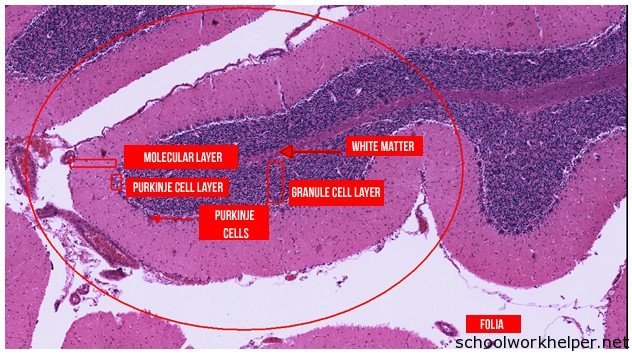
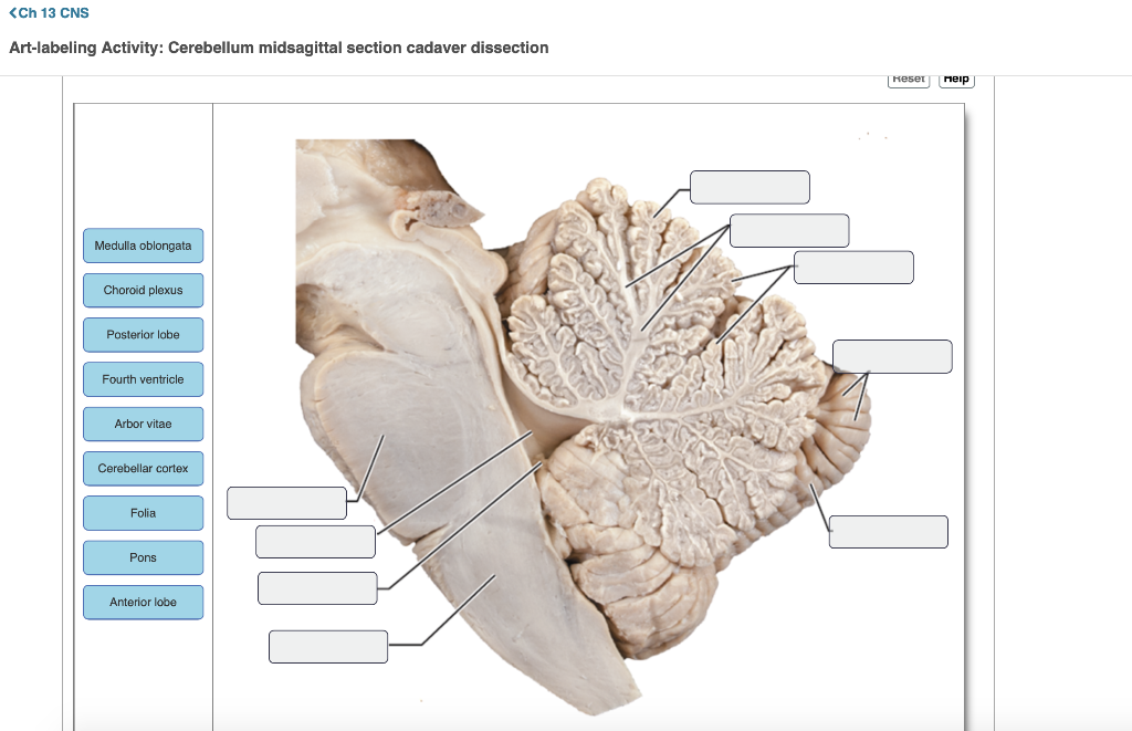
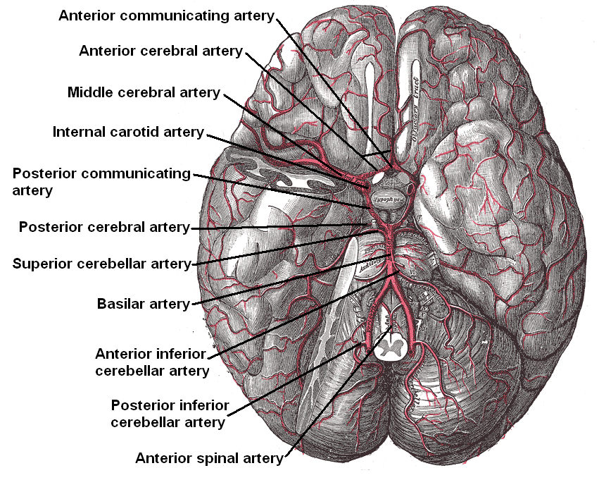
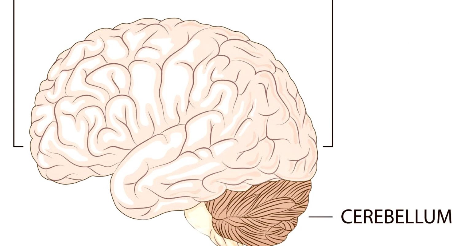
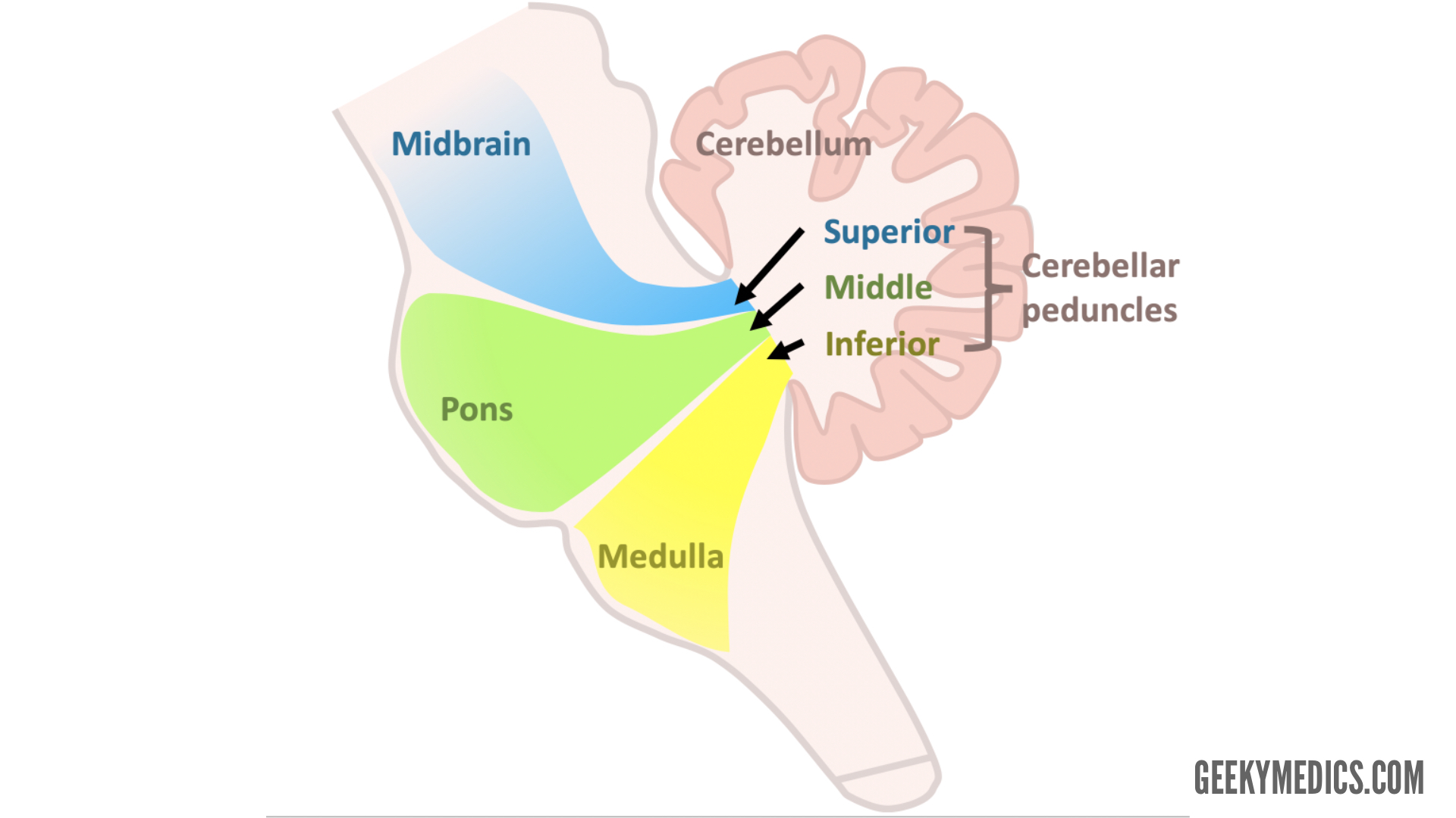
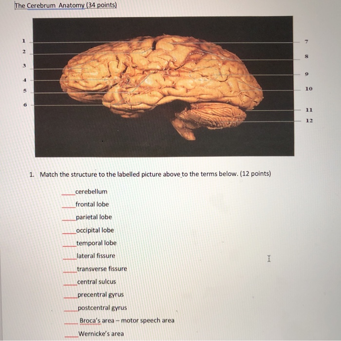
42 cerebellum labelled
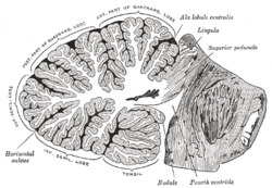
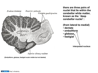
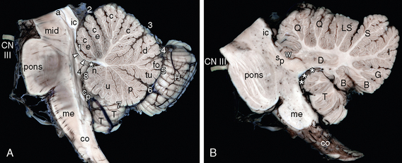
en.wikipedia.org › wiki › Arbor_vitae_(anatomy)Arbor vitae (anatomy) - Wikipedia It brings sensory and motor information to and from the cerebellum. The arbor vitae is located deep in the cerebellum. Situated within the arbor vitae are the deep cerebellar nuclei; the dentate, globose, emboliform and the fastigial nuclei. These four different structures lead to the efferent projections of the cerebellum. The Breasts - Structure - Vasculature - TeachMeAnatomy Feb 07, 2022 · The breasts are paired structures located on the anterior thoracic wall, in the pectoral region. They are present in both males and females, yet are more prominent in females following puberty. In females, the breasts contain the mammary glands – an accessory gland of the female reproductive system. The mammary glands are the key structures involved in …
Greater Trochanter - Location, Functions, Anatomy, Diagrams The greater trochanter is a bony protrusion located in the upper extremity, or femur epiphysis. The greater trochanter is located at the junction between the neck and the shaft of the femur bone.
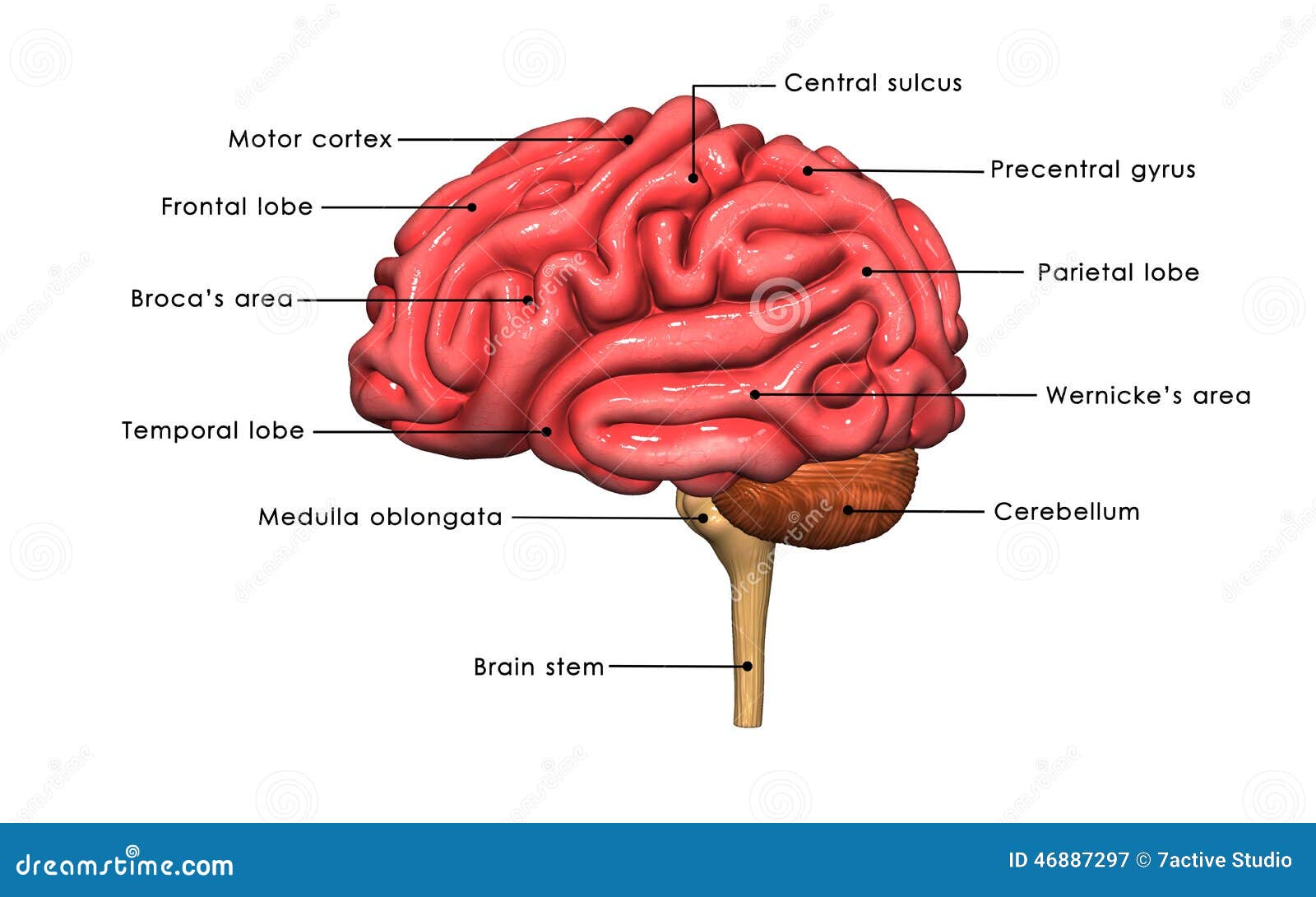
Cerebellum labelled
en.wikipedia.org › wiki › Brain_cellBrain cell - Wikipedia Brain cells make up the functional tissue of the brain.The rest of the brain tissue is structural or connective called the stroma which includes blood vessels.The two main types of cells in the brain are neurons, also known as nerve cells, and glial cells also known as neuroglia. en.wikipedia.org › wiki › Stellate_cellStellate cell - Wikipedia Stellate cells are derived from dividing progenitors in the white matter of postnatal cerebellum. Dendritic trees can vary between neurons. There are two types of dendritic trees in the cerebral cortex, which include pyramidal cells , which are pyramid shaped and stellate cells which are star shaped. Stellate cell - Wikipedia Stellate cells are any neuron in the central nervous system that have a star-like shape formed by dendritic processes radiating from the cell body. Many Stellate cells are GABAergic and are located in the molecular layer of the cerebellum. Stellate cells are derived from dividing progenitors in the white matter of postnatal cerebellum. Dendritic trees can vary between …
Cerebellum labelled. Thenar muscles: Anatomy, innervation and function | Kenhub Jul 22, 2022 · The flexor pollicis brevis is the most medial of the thenar muscles. It arises by two muscle heads (superficial and deep) which are separated by the tendon of flexor pollicis longus.The superficial head originates from the flexor retinaculum and the tubercle of the trapezium bone, while the deep head originates from the trapezoid and capitate bones. . From … fsl.fmrib.ox.ac.uk › fsl › fslwikiAtlases - FslWiki - University of Oxford A single subject's structural image was hand segmented, and the labels were then propagated to more than 50 subjects' structural images using nonlinear registration. Each resulting labelled brain was then transformed into MNI152 space using affine registration, before averaging segmentations across subjects to produce the final probability images. Page: American Journal of Ophthalmology Mar 03, 2022 · The American Journal of Ophthalmology is a peer-reviewed, scientific publication that welcomes the submission of original, previously unpublished manuscripts directed to ophthalmologists and visual science specialists describing clinical investigations, clinical observations, and clinically relevant laboratory investigations. Medial and lateral pterygoid muscle: Anatomy and function | Kenhub Apr 26, 2022 · Pterygoid muscles. The pterygoid muscles are two of the four muscles of mastication, located in the infratemporal fossa of the skull.These muscles are: lateral pterygoid and medial pterygoid. The primary function of the pterygoid muscles is to produce movements of the mandible at the temporomandibular joint.Both muscles are innervated by branches of the …
Global profiling of miRNAs and the hairpin precursors: insights … Feb 08, 2013 · Total RNA was isolated from adult mouse tissues (cerebellum, cortex, heart, kidney, liver, lung, ovary, skeletal muscle, ... 32 P-labelled CandidateMMU32 pre-miRNA (lane 3–5) and CandidateMMU33 pre-miRNA (lane 8–10) were incubated with rDicer for an increasing time. Lane 1 and 6 showed the size marker. Arbor vitae (anatomy) - Wikipedia The arbor vitae / ˌ ɑːr b ɔːr ˈ v aɪ t iː / (Latin for "tree of life") is the cerebellar white matter, so called for its branched, tree-like appearance.In some ways it more resembles a fern and is present in both cerebellar hemispheres. It brings sensory and motor information to and from the cerebellum.The arbor vitae is located deep in the cerebellum. Tutorials/LabelFreeSurfer - Brainstorm - University of Southern … Aug 09, 2022 · Import the sub-cortical atlas *aseg.mgz as volume atlases and labelled surfaces ; Import the cortical thickness maps ; The files you can see in the database explorer at the end (with Volume atlases + Thickness): MRI: The T1 MRI of the subject, imported from the MGH file format (T1.mgz) Volume parcellations: *aseg.mgz, see tutorial Explore the ... › test-catalog › overviewENC2 - Overview: Encephalopathy, Autoimmune/Paraneoplastic ... Strips are washed to remove unbound antibodies and then incubated with anti-human IgG antibodies (alkaline phosphatase-labelled) for 30 minutes. The strips are again washed to remove unbound anti-human IgG antibodies and nitroblue tetrazolium chloride/5-bromo-4-chloro-3-indolylphosphate (NBT/BCIP) substrate is added.
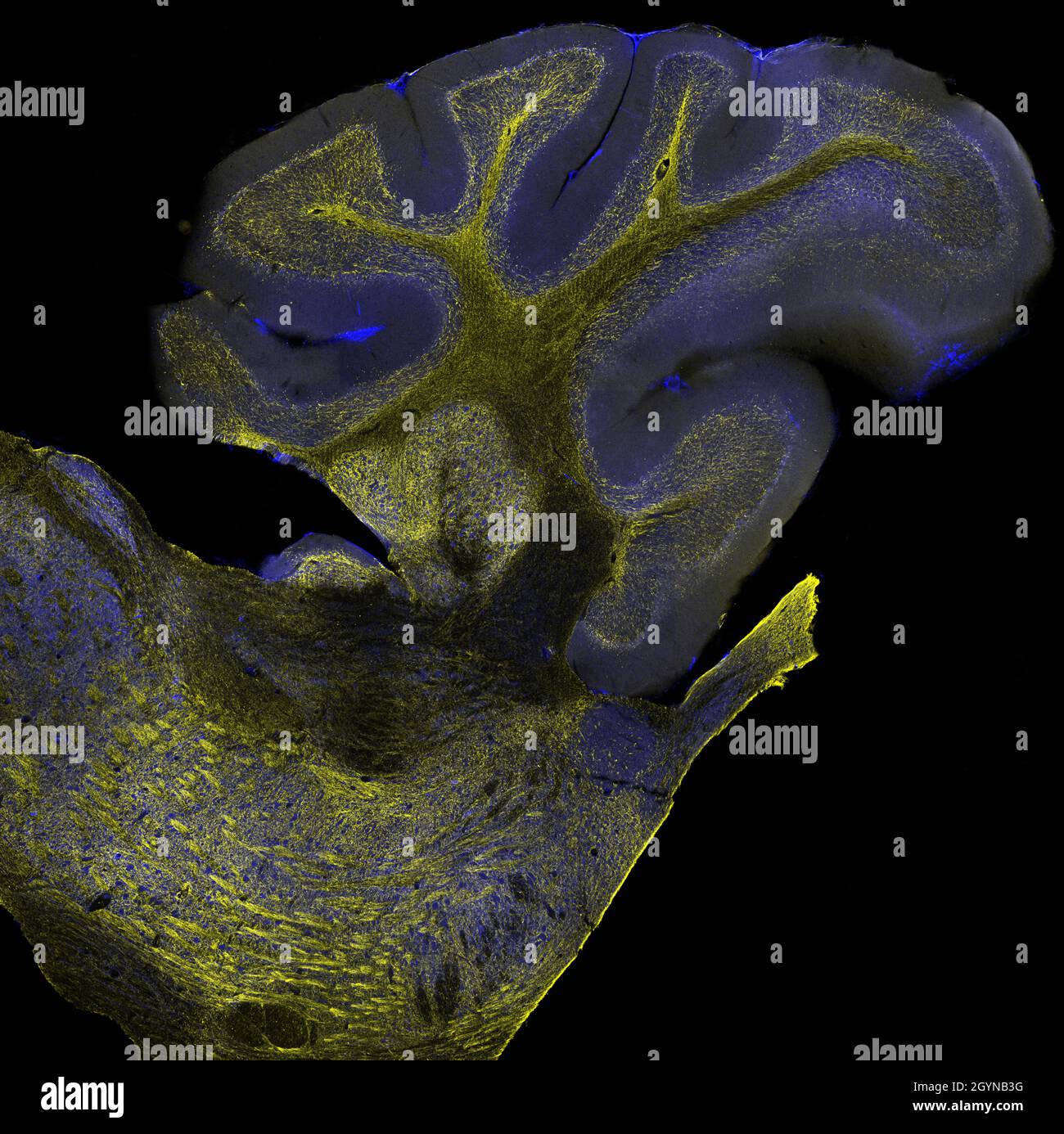
Transcriptomic mapping uncovers Purkinje neuron plasticity … May 11, 2022 · f, RNA ISH analyses using fluorescence-labelled Aldoc or Plcb4 RNA probes and probes for newly defined Purkinje neuron subtypes (Sv2c and Sorcs2). Scale bars, 800 μm (left) and 200 μm (right). ZEISS LSM 980 with Airyscan 2 Mouse brain cerebellum labelled with anti-calbinding (Alexa-568) and anti-GFAP (Alexa-488). The fluorophores were both excited with the two-photon laser at 780 nm and the emission spectra were simultaneously collected by the BIG.2 detector. 3D Tilling and Stitching were used to cover whole structure, and an orthogonal projection was created in ... en.wikipedia.org › wiki › Grey_matterGrey matter - Wikipedia Grey matter is distributed at the surface of the cerebral hemispheres (cerebral cortex) and of the cerebellum (cerebellar cortex), as well as in the depths of the cerebrum (the thalamus; hypothalamus; subthalamus, basal ganglia – putamen, globus pallidus and nucleus accumbens; as well as the septal nuclei), cerebellum (deep cerebellar nuclei ... Stellate cell - Wikipedia Stellate cells are any neuron in the central nervous system that have a star-like shape formed by dendritic processes radiating from the cell body. Many Stellate cells are GABAergic and are located in the molecular layer of the cerebellum. Stellate cells are derived from dividing progenitors in the white matter of postnatal cerebellum. Dendritic trees can vary between …
en.wikipedia.org › wiki › Stellate_cellStellate cell - Wikipedia Stellate cells are derived from dividing progenitors in the white matter of postnatal cerebellum. Dendritic trees can vary between neurons. There are two types of dendritic trees in the cerebral cortex, which include pyramidal cells , which are pyramid shaped and stellate cells which are star shaped.
en.wikipedia.org › wiki › Brain_cellBrain cell - Wikipedia Brain cells make up the functional tissue of the brain.The rest of the brain tissue is structural or connective called the stroma which includes blood vessels.The two main types of cells in the brain are neurons, also known as nerve cells, and glial cells also known as neuroglia.




:background_color(FFFFFF):format(jpeg)/images/library/10113/structure-of-cerebellum_english.jpg)

:watermark(/images/watermark_only.png,0,0,0):watermark(/images/logo_url.png,-10,-10,0):format(jpeg)/images/anatomy_term/uvula-vermis/0WItsn7kVfwPmmQ7TSSOA_Uvula_01.png)







:background_color(FFFFFF):format(jpeg)/images/library/11646/basic-anatomy-of-the-brain_english_copy.jpeg)


/profile-of-man-s-head-with-brain-anatomy-labeled-on-white-background-1093597090-f6a5470b98a4453997931b1cb72fb47d.jpg)
Post a Comment for "42 cerebellum labelled"