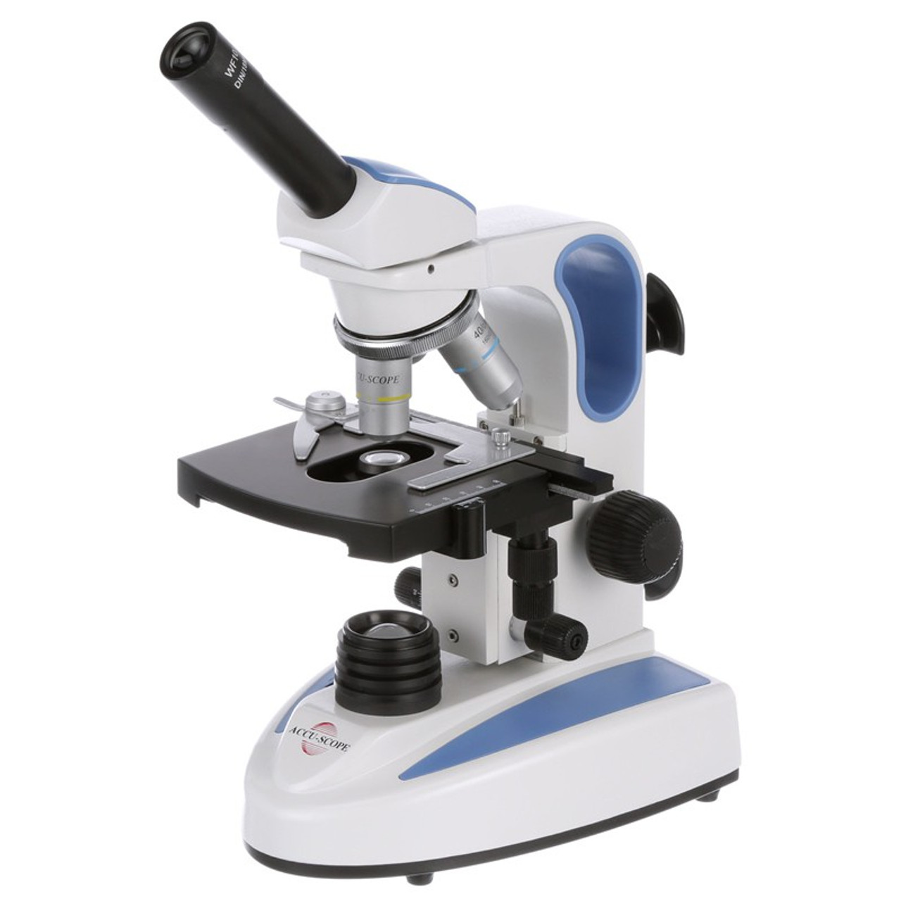
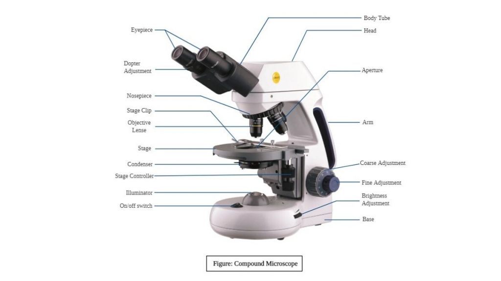
39 labelled diagram of compound microscope
› pdf › ieFor Objective Questions and NCERT Solutions ... microscope. The particles are visible under microscope. (iv) The particle of a true solution can be recovered. The particles of a colloidal solution cannot be recovered. (v) The particles of a true solution do not scatter light. The particles of a colloidal solution scatter light. 49. Explain sublimation process with labelled diagram. Ans : cbseacademic.nic.in › web_material › SQPClass-X Science-086 SAMPLE QUESTION PAPER-19 TIME: 3 ... - CBSE 17. A compound A (C 2H 4O 2) reacts with Na metal to form a compound ‘B’ and evolves a gas which burns with a pop sound. Compound ‘A’ on treatment with an alcohol ‘C’ in presence of an acid forms a sweet smelling compound ‘D’ (C 4H 8O 2). On addition of NaOH to ‘D’ gives back B and C. Identify A, B, C and D write the ...
microbenotes.com › under-the-microscopeAmazing 27 Things Under The Microscope With Diagrams May 13, 2022 · Observation under the compound microscope. Under a compound microscope, the differences between the sand particles become more apparent. It is visible that the shape, size color, and texture of individual particles vary within the sand collected from the same place. Some grains might appear smooth, while others appear irregular and sharp.
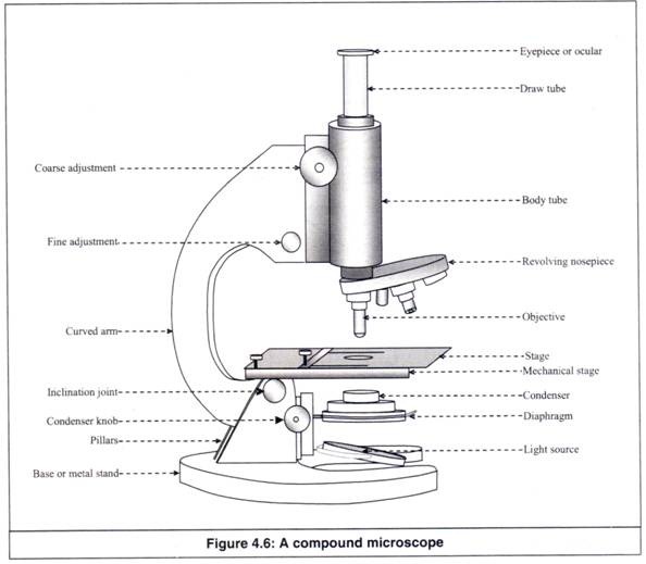
Labelled diagram of compound microscope
› cell-biologyTop 16 Techniques Used in Cell Biology (With Diagram) ADVERTISEMENTS: The following points highlight the top sixteen techniques used in cell biology. Some of the techniques are: 1. Immunofluorescence Microscopy 2. Ion-Exchange Chromatography 3. Affinity Chromatography 4. Partition and Adsorption Chromatography 5. Gel Filtration Chromatography 6. Radioactive Tracer Technique 7. Radioimmunoassay (RIA) 8. Enzyme Immunoassay 9. Spectroscopy and ... microscopewiki.com › compound-microscopeCompound Microscope – Diagram (Parts labelled), Principle and ... Feb 03, 2022 · See: Labeled Diagram showing differences between compound and simple microscope parts Structural Components. The three structural components include. 1. Head. This is the upper part of the microscope that houses the optical parts en.wikipedia.org › wiki › EyeEye - Wikipedia Photoreception is phylogenetically very old, with various theories of phylogenesis. The common origin of all animal eyes is now widely accepted as fact.This is based upon the shared genetic features of all eyes; that is, all modern eyes, varied as they are, have their origins in a proto-eye believed to have evolved some 650-600 million years ago, and the PAX6 gene is considered a key factor in ...
Labelled diagram of compound microscope. › 39605905 › Cambridge_CheckpointCambridge Checkpoint Science Coursebook 8 - Academia.edu This captivating Coursebook provides coverage of stage 8 of the revised Cambridge Secondary 1 curriculum framework. It is endorsed by Cambridge International Examinations for use with their programme. The series is written by a highly experienced en.wikipedia.org › wiki › EyeEye - Wikipedia Photoreception is phylogenetically very old, with various theories of phylogenesis. The common origin of all animal eyes is now widely accepted as fact.This is based upon the shared genetic features of all eyes; that is, all modern eyes, varied as they are, have their origins in a proto-eye believed to have evolved some 650-600 million years ago, and the PAX6 gene is considered a key factor in ... microscopewiki.com › compound-microscopeCompound Microscope – Diagram (Parts labelled), Principle and ... Feb 03, 2022 · See: Labeled Diagram showing differences between compound and simple microscope parts Structural Components. The three structural components include. 1. Head. This is the upper part of the microscope that houses the optical parts › cell-biologyTop 16 Techniques Used in Cell Biology (With Diagram) ADVERTISEMENTS: The following points highlight the top sixteen techniques used in cell biology. Some of the techniques are: 1. Immunofluorescence Microscopy 2. Ion-Exchange Chromatography 3. Affinity Chromatography 4. Partition and Adsorption Chromatography 5. Gel Filtration Chromatography 6. Radioactive Tracer Technique 7. Radioimmunoassay (RIA) 8. Enzyme Immunoassay 9. Spectroscopy and ...
Compound Microscope Review - ppt download
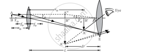
Draw a Labelled Ray Diagram Showing the Formation of a Final ...

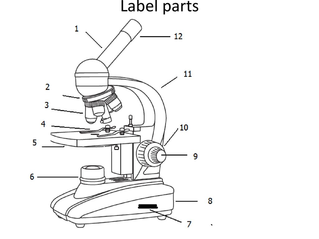
Parts of a Microscope Microscope Basics. Label the Compound ...

BIOLOGY FROM 1 | EQUIPMENTS USED FOR OBSERVATION | Cours ...

Biology - labeling a compound microscope Diagram | Quizlet
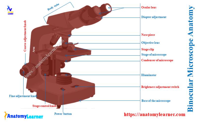
Binocular Microscope Anatomy - Parts and Functions with a ...

Labeling the Parts of the Microscope | Microscope activity ...

label microscope diagram | Charts | Microscope, Anatomy bones ...

Microscope Diagram Labeled, Unlabeled and Blank | Parts of a ...

General Biology | Carlson Stock Art | General biology ...
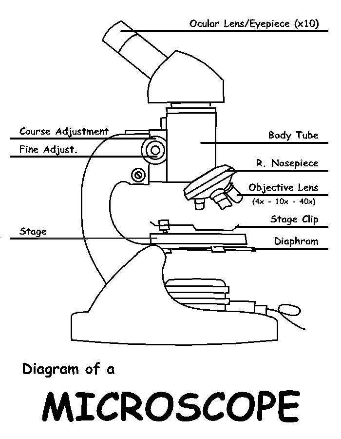
Diagram of a Microscope by ScienceDoodles on DeviantArt
Compound Microscope Parts, Functions, and Labeled Diagram ...
Compound Microscope Parts, Diagram Definition, Application ...

Label Microscope Diagram - EnchantedLearning.com
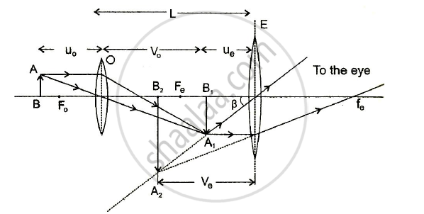
Draw a Labelled Ray Diagram Showing the Formation of a Final ...

Draw a well labelled diagram of a microscope. - Brainly.in
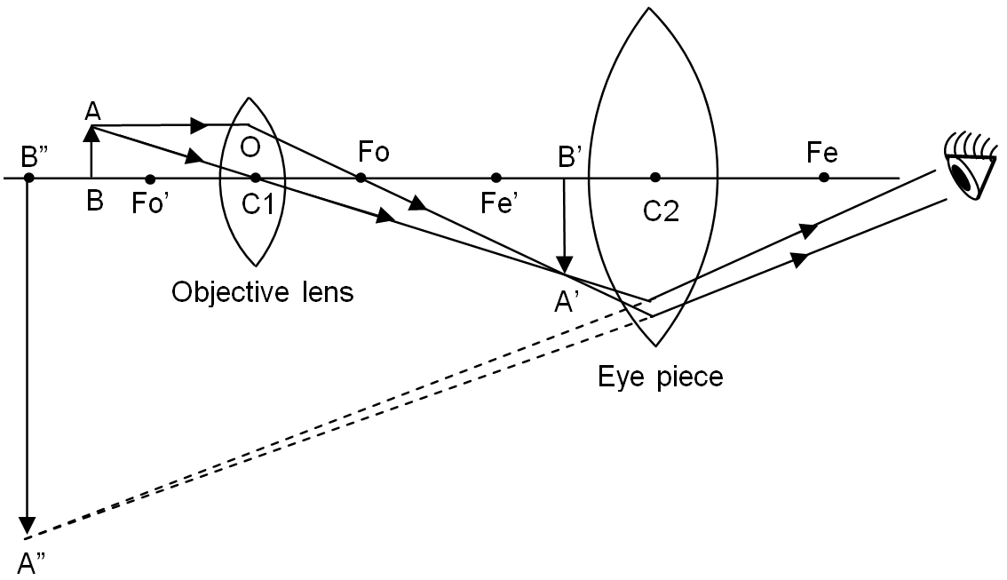
Draw a labelled ray diagram of a compound microscope and ...

AmScope 40X-2000X 3W LED Siedentopf Trinocular Compound ...

Parts of a Compound Microscope and Their Functions
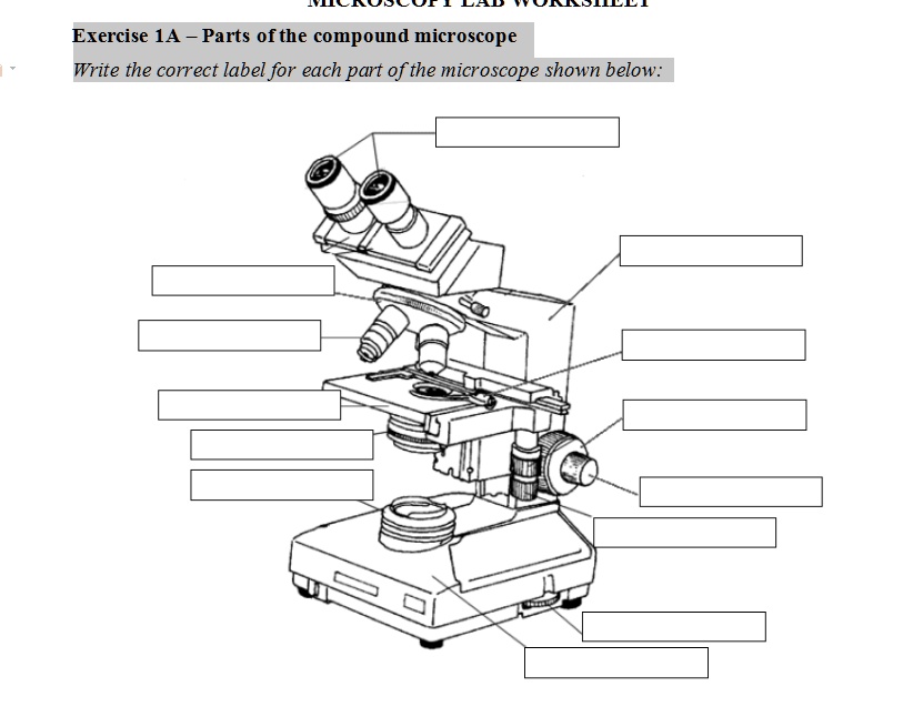
SOLVED: Exercise 1A _ Parts ofthe compound microscope Write ...

Biology 4 U on Twitter: "Try this labelled diagram Quiz on ...
The Compound Light Microscope Label the following parts on ...
What is a diagram of a plant and animal cell under an ...

Draw a neat labelled diagram of a compound microscope and ...

Diagram of a Compound Microscope

Parts of a microscope with functions and labeled diagram
parts of microscope with diagram - Clip Art Library

2.1 " Compound Microscope" | Download Scientific Diagram

Difference between Simple and Compound Microscope ...
The Compound Light Microscope Label the following parts on ...

Identify the parts labelled A and B.
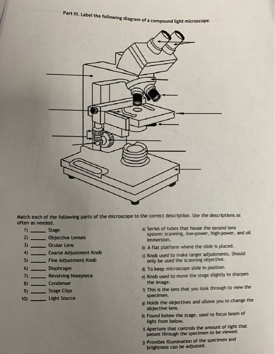
Solved Part III. Label the following diagram of a compound ...
Microscope Types (with labeled diagrams) and Functions

Compound Microscope: Parts of Compound Microscope

a) Explain the working of a compound microscope with the help ...
Label a microscope - Teaching resources

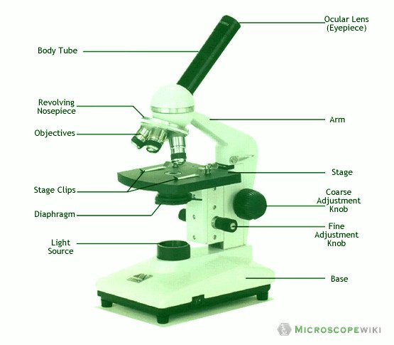
Microscope, Microscope Parts, Labeled Diagram, and Functions

Microscope Parts and Functions

compound microscope Diagram | Quizlet
Post a Comment for "39 labelled diagram of compound microscope"